Veterinary ultrasound equipment has become one of the most relied-upon diagnostic assets in modern animal healthcare. Whether used for abdominal imaging, cardiac assessment, or reproductive evaluation, ultrasound systems give clinicians real-time visibility into animal anatomy without radiation exposure or invasive procedures. Selecting the right unit, however, is not a matter of brand preference—it’s a strategic, operational decision that influences diagnostic accuracy, workflow speed, and long-term return on investment.
This framework provides a clear, factual method for evaluating veterinary ultrasound systems based on measurable criteria: system type, imaging performance, probe ecosystem, workflow integration, maintenance requirements, and cost of ownership. Each section outlines what professionals should verify before purchase, ensuring that every investment aligns with the practice’s operational objectives and patient mix.
The Core Purpose of Veterinary Ultrasound Equipment
Ultrasound imaging enables clinicians to view organs, tissues, and vascular structures in real time—making it a cornerstone of diagnostics for companion, equine, and livestock medicine.
Veterinary ultrasound works by transmitting high-frequency sound waves through tissue and converting the returning echoes into visual images. Its non-invasive nature allows repeat scanning without risk, supporting early detection, treatment monitoring, and post-operative assessment. A properly configured system supports multiple anatomical regions, from abdominal organs and reproductive systems to cardiac chambers and musculoskeletal structures.
Because animal species differ widely in size and anatomy, equipment selection directly determines image penetration, resolution, and diagnostic reliability. Inconsistent performance across species often reflects a mismatch between probe frequency, transducer design, and system processing capability. Recognizing these variables is the first step toward objective equipment evaluation.
Main Categories of Veterinary Ultrasound Equipment
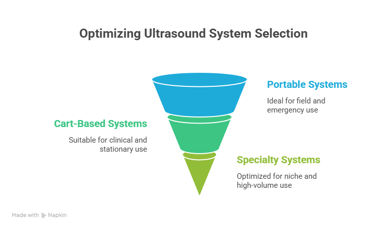
Veterinary ultrasound systems fall into three categories—portable, cart-based, and specialty units—each suited to distinct environments and workloads.
- Portable Ultrasound Systems
Compact and battery-powered, portable units are built for mobility. They are essential for ambulatory veterinarians, mixed-animal practitioners, and emergency responders. Typical configurations include a lightweight console (under 15 lb), 2-3 probe ports, and internal battery life of up to four hours. Their main strengths are field readiness and convenience; their trade-offs include smaller display size and limited advanced imaging modes.
- Cart-Based Ultrasound Systems
Designed for clinical settings, these systems offer higher processing power, advanced Doppler modes, and superior image quality. They accommodate multiple probes simultaneously and integrate seamlessly with hospital IT infrastructure. The larger footprint supports extended use but limits mobility—making them ideal for stationary diagnostic rooms and referral centers.
- Specialty Ultrasound Systems
Purpose-built for niche applications such as cardiology, reproduction, or orthopedic imaging, specialty systems deliver precision and depth through customized probes and software packages. They are optimized for specific anatomical studies rather than general practice. Facilities that handle high imaging volume or referral-level diagnostics benefit most from this configuration.
Understanding these categories helps align equipment specifications with operational goals—whether mobility, throughput, or specialization.
Technical Specifications That Determine Image Quality
The clarity and diagnostic reliability of ultrasound images depend on transducer frequency, signal processing, and frame-rate stability.
Transducer Frequency:
Lower frequencies (2–5 MHz) penetrate deeper tissue, making them suitable for large animals. Higher frequencies (7–15 MHz) provide finer resolution for small animals and superficial structures. Clinics serving a wide species range require systems supporting multiple frequency bands to ensure diagnostic flexibility.
Signal Processing and Dynamic Range:
A broader dynamic range captures subtle tissue contrasts, crucial for distinguishing lesions or vascular details. Systems with advanced beam-forming and adaptive image processing deliver higher fidelity under varied scanning conditions.
Frame Rate and Refresh Stability:
Consistent frame rates are essential for moving structures such as the heart or gastrointestinal tract. Dropped frames or latency can obscure motion-related findings, reducing diagnostic accuracy.
Display and Interface:
A high-resolution, anti-glare monitor and intuitive control layout reduce operator fatigue. Adjustable brightness and ergonomic controls improve efficiency during long imaging sessions.
Image quality is therefore not a single specification—it’s the combined outcome of transducer design, electronics, and operator interface.
Operational Considerations When Selecting Equipment
Evaluate usability, durability, and service access alongside imaging specifications to ensure reliability across daily operations.
Portability and Power:
Field veterinarians should confirm battery life, recharge time, and total operating hours per cycle. Systems designed for mobile use often feature shock-resistant housings and sealed keyboards to prevent dust or moisture intrusion.
User Interface Design:
Touchscreen controls allow faster adjustments but may be less precise when wearing gloves. Physical buttons provide tactile feedback and durability. Assess which configuration best suits your staff’s workflow and environment.
Maintenance Accessibility:
Simplified housings with easy-to-remove panels facilitate internal cleaning and component inspection. Consider the manufacturer’s maintenance intervals and whether calibration can be performed in-house or requires shipping.
Warranty and Support:
Review service terms carefully. Standard coverage ranges from one to five years, often excluding probes. Extended coverage may reduce long-term costs if probe replacement or software upgrades are included.
Evaluating the Probe Ecosystem
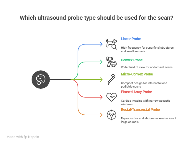
The probe selection defines the system’s diagnostic range and determines how effectively a clinic can serve varied patients.
Probe Types:
- Linear: High frequency for tendons, superficial structures, and small animals.
- Convex: Wider field of view for abdominal and general scans.
- Micro-Convex: Compact design ideal for intercostal and pediatric scans.
- Phased Array: Used for cardiac imaging where narrow acoustic windows are common.
- Rectal/Transrectal: Designed for large-animal reproductive and abdominal evaluations.
Compatibility:
Systems supporting multi-probe ports enable rapid switching without reconfiguration. Evaluate connector durability and cable flexibility, especially for portable setups where frequent plugging occurs.
Lifecycle:
Probe longevity depends on handling, disinfection chemicals, and mechanical stress. Establish a replacement schedule and inspect cables for strain relief and wear. A probe’s average lifespan ranges from three to five years under normal use.
Cleaning and Storage:
Dedicated probe holders and temperature-controlled storage reduce mechanical stress. Follow manufacturer-approved disinfectants to maintain membrane integrity.
Workflow Integration and Data Management
Efficient data handling is vital for clinical documentation, collaboration, and compliance.
DICOM Compatibility:
Ensure the system supports DICOM protocols for standardized data exchange with imaging archives or referral centers. This enables secure storage, review, and transfer of diagnostic images.
Internal Storage and Export:
Typical configurations range from 8 GB internal memory to multi-terabyte network drives. Systems should allow USB and Ethernet export, plus direct wireless transfer where available.
Software Tools:
Integrated measurement packages streamline reporting. Systems with anatomical labeling, trace functions, and customizable templates reduce clerical time and enhance record accuracy.
Practice Management Integration:
Interfacing with patient databases ensures each scan is automatically linked to medical records, preventing misfiled images. Evaluate compatibility with your clinic’s existing software infrastructure before purchase.
Maintenance, Calibration, and Service Life
Routine maintenance preserves accuracy, uptime, and hygiene—directly affecting the lifespan of your investment.
Cleaning Procedures:
After every use, probes must be wiped with approved enzymatic or alcohol-free solutions. The console surface should be cleaned with non-abrasive cloths to prevent residue buildup.
Calibration and Performance Checks:
At least annually, verify calibration for time-gain compensation, grayscale linearity, and probe sensitivity. Many systems include built-in diagnostic tests that simplify verification.
Software Updates:
Maintain up-to-date firmware to benefit from performance enhancements and bug fixes. Updates should not affect stored images; confirm automatic backup before installing patches.
Service Life Expectancy:
A high-quality system typically provides 8–10 years of service with routine maintenance. Systems used heavily in field environments may require probe replacement sooner due to mechanical stress.
Cost-of-Ownership Considerations:
Factor recurring costs such as gel, disinfectants, and calibration service into annual budgets. Preventive maintenance is more cost-effective than emergency repair.
Pricing and Budget Allocation Framework
A balanced budget must account for purchase price, accessories, and operational continuity.
Typical Price Ranges:
- Entry-Level Portable Systems: CAD $1,500 – $4,000
- Mid-Range Cart-Based Systems: CAD $6,000 – $15,000
- Premium Specialty Systems: CAD $20,000 and above
Pricing varies by included probes, imaging modes, and warranty coverage. When comparing units, always confirm the number of probes bundled, display resolution, and after-sale service terms.
Accessory Costs:
Include replacement cables, battery packs, cleaning solutions, and protective cases. For high-volume use, budgeting 5 – 10 percent of the equipment cost annually for consumables and maintenance is prudent.
Service Contracts:
Extended service contracts typically range from 8 – 12 percent of purchase cost per year. They may include loaner units during repair, reducing operational downtime.
Depreciation Planning:
Straight-line depreciation over seven years is standard in most clinics. Properly maintained systems often retain 20 – 30 percent of their original value for trade-in toward upgrades.
Checklist for Purchasing Veterinary Ultrasound Equipment
A concise verification list ensures that technical and operational requirements are both satisfied before purchase.
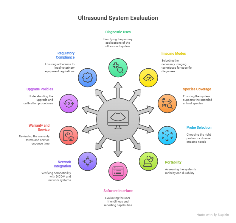
Pre-Purchase Checklist
- Define primary diagnostic uses: abdominal, cardiac, reproductive, or musculoskeletal.
- Determine required imaging modes: B-mode, M-mode, color Doppler, power Doppler.
- Confirm species coverage and transducer frequency range.
- Evaluate number and type of probes supplied.
- Verify portability requirements: weight, battery duration, shock resistance.
- Assess software interface for ease of use and reporting functionality.
- Check for DICOM and network integration.
- Review warranty length, coverage limits, and service response time.
- Examine upgrade and calibration policies.
- Ensure compliance with local veterinary equipment regulations.
Completing this checklist before procurement avoids costly oversights and ensures the unit integrates seamlessly into daily operations.
Common Mistakes Clinics Should Avoid
Missteps in equipment selection and upkeep often result in hidden costs and diagnostic inefficiencies.
- Choosing on Price Alone: Low-cost units may limit probe options or lack advanced modes, forcing premature replacement.
- Overlooking Maintenance: Neglecting calibration or probe inspection shortens lifespan and increases downtime.
- Ignoring Workflow Fit: Systems without data integration create manual workarounds and risk data loss.
- Insufficient Training: Even advanced systems produce poor results if staff are unfamiliar with presets and gain controls.
- Lack of Environmental Planning: Units without dust or moisture protection fail early in mobile or farm conditions.
Avoiding these errors ensures consistent imaging performance and protects the financial investment over time.
FAQs
How does veterinary ultrasound equipment differ from human ultrasound machines?
Veterinary systems are built for anatomical diversity, offering wider frequency ranges and specialized probes for different species. They include presets for dogs, cats, horses, and livestock, whereas human systems are optimized for uniform anatomy and lack species-specific imaging software.
What are the essential features to look for in vet ultrasound equipment?
Key features include high-resolution imaging, multiple probe ports, advanced Doppler modes, durable construction, and DICOM compatibility. Ease of use, quick boot time, and efficient data export functions are equally important for daily clinical operations.
Which imaging modes are standard in modern veterinary ultrasound systems?
Most systems offer B-mode for structure visualization, M-mode for motion assessment, and Color or Power Doppler for blood flow analysis. Higher-end systems add Harmonic Imaging and 3D/4D reconstruction for detailed studies.
Can portable ultrasound machines match the performance of cart-based units?
Portable systems have improved significantly and can deliver comparable image quality for general and reproductive exams. However, cart-based systems maintain an advantage in complex imaging tasks like advanced cardiac or vascular diagnostics.
What factors determine the right probe selection for my clinic?
Probe choice depends on animal size, anatomy, and procedure type. Linear probes excel for small-animal and tendon imaging, convex probes for abdominal work, and rectal probes for large-animal reproduction. A mixed-practice clinic should maintain at least three probe types.
How long do veterinary ultrasound probes typically last?
With proper care and approved disinfectants, probes last between three and five years. Frequent cable bending or use of harsh cleaning agents can shorten this lifespan considerably.
Is ultrasound training necessary for veterinarians and technicians?
Yes. Even the best equipment underperforms without trained operators. Training improves diagnostic accuracy, shortens scan times, and reduces repeat examinations, improving both workflow and patient comfort.
How should veterinary ultrasound machines be cleaned and maintained?
Follow manufacturer guidelines strictly. Probes should be cleaned with non-abrasive enzymatic solutions immediately after use, and the system should be covered when idle. Schedule annual calibration checks to maintain consistent image quality.
What is the average cost of vet ultrasound equipment in Canada?
Entry-level portable systems start around CAD $1,500–$3,000, mid-range units range from CAD $6,000–$12,000, and premium systems can exceed CAD $20,000 depending on configuration, probe count, and Doppler capability.
How does warranty length affect total cost of ownership?
Longer warranties reduce lifetime costs by covering probe repair and software updates. A five-year warranty can offset the expense of at least one major component replacement.
Can ultrasound machines be upgraded after purchase?
Most modern systems allow software updates and optional probe additions. Verify that your unit supports modular upgrades to extend usability without replacing the main console.
How much image storage is recommended for busy clinics?
A minimum of 500 GB is advisable for high-volume clinics. Systems should also support USB or network backups to prevent data loss and simplify long-term record management.
What are signs that my ultrasound machine needs servicing?
Indicators include image distortion, inconsistent gain response, or unresponsive controls. Regular calibration can identify these issues early and prevent downtime.
Do all veterinary ultrasound machines support DICOM integration?
No. Lower-priced systems may rely solely on USB export. For clinics with digital archives, DICOM compatibility is essential for seamless storage and retrieval.
How does ultrasound contribute to practice growth?
Offering in-house imaging increases diagnostic efficiency, patient retention, and service revenue. Clinics that integrate ultrasound often see higher case acceptance and fewer external referrals.
Making the Right Investment in Veterinary Ultrasound Equipment
Choosing the right ultrasound system isn’t just a technical purchase—it’s a long-term investment in diagnostic precision, workflow reliability, and client confidence.
Veterinary ultrasound equipment has become an indispensable diagnostic tool across small-animal, equine, and mixed-practice settings. The difference between a reliable asset and a costly underperformer lies in how carefully it’s evaluated before purchase. Clinics that define their imaging requirements, compare probe ecosystems, and budget for maintenance achieve longer service life and better clinical outcomes.
When comparing systems, prioritize clarity of imaging, multi-species adaptability, and the ability to integrate smoothly with your existing digital records. Verify service support and probe durability before finalizing any purchase. Maintenance and training should be viewed not as optional add-ons but as integral components of your investment strategy.
BOMImed provides access to a curated range of veterinary ultrasound systems engineered for consistent performance, field readiness, and long-term support. Each unit is built for accuracy, durability, and ease of integration within modern veterinary environments.
Explore BOMImed’s Vet Ultrasound Equipment collection to find the system that matches your clinic’s needs—ensuring that every scan delivers clarity, confidence, and clinical precision.