The Purpose of a Clinical-First Buying Framework
Veterinarians often purchase ultrasound equipment by comparing specifications, pricing tiers, or general categories. A clinical-first framework reverses that sequence by anchoring the decision in the realities of patient care, workflow pacing, diagnostic demand, and long-term practice strategy. This approach prevents overbuying, underbuying, or choosing systems mismatched to actual case patterns.
A clinical-first evaluation method aligns the machine’s capabilities with the type of medicine the practice delivers every day. This produces more consistent imaging results, higher utilization rates, and a clearer return on investment. It also ensures that the equipment remains relevant as the practice expands services or brings new specialties in-house.
When equipment decisions are guided by case profiles instead of marketing claims, veterinarians gain stronger diagnostic confidence and fewer operational limitations. It becomes easier to select systems that perform reliably during routine exams, emergency triage, and advanced specialty work without compromising the overall workflow.
Identifying the Clinical Demands Driving the Veterinary Equipment Decision
Understanding the Practice’s Diagnostic Profile
Every practice develops recognizable diagnostic patterns based on species, case complexity, patient volume, and clinician expertise. These patterns define how ultrasound is used throughout the day and set the baseline for equipment requirements. A machine that excels in small-animal abdominal imaging may struggle in equine tendon assessment, while a system optimized for reproductive work may not provide the resolution needed for internal medicine.
Clinicians benefit from mapping out their most frequent scan types and identifying those they intend to expand. This creates a practical list of imaging demands that the ultrasound machine must consistently meet. When these needs are organized clearly, the rest of the buying decision becomes more straightforward.
Certain practices use ultrasound as a broad screening tool, while others use it as a precision instrument for cardiac, soft-tissue, or orthopedic evaluation. Understanding where the practice falls on that spectrum determines how much emphasis to place on transducer selection, Doppler modes, penetration depth, and workstation ergonomics.
Evaluating Case Complexity and Specialty Needs
A general practice performing weekly abdominal scans has different needs from a specialty clinic conducting advanced echocardiography or oncology assessments. Matching equipment capabilities to projected case complexity ensures that the system remains productive even as the practice evolves.
Practices offering reproductive services require consistent image penetration and customizable measurement tools. Clinics managing chronic cardiac cases need reliable Doppler performance and stable frame rates. Veterinary teams working with exotics or small mammals require fine-detail resolution and precise focal control.
The objective is to identify the minimum clinical threshold the equipment must meet. Once these standards are defined, the decision becomes less about abstract features and more about whether the machine fulfills the practice’s real diagnostic workload.
Determining the Role of Ultrasound in Emergency and Point-of-Care Situations
Ultrasound systems used during emergency triage must be immediately accessible, fast to boot, and simple to position. Practices offering after-hours care or rapid-response services require a machine capable of delivering clear images without lengthy adjustments.
Point-of-care use often favors systems with intuitive interfaces, durable housings, and transducers that remain accurate under fast-paced conditions. In emergency contexts, clinicians rely on the machine’s ability to deliver clear organ boundaries, free fluid identification, and stable Doppler signals without multiple passes.
Establishing the degree of emergency usage influences decisions on portability, cable durability, and the system’s overall resilience in high-pressure environments.
Matching Equipment Specifications to Clinical Requirements
Prioritizing Image Quality
General image quality is not a single specification but the product of resolution, contrast, depth penetration, dynamic range, and artifact control. A clinical-first approach evaluates these elements based on real diagnostic demands rather than theoretical maximums.
Soft-tissue evaluation requires smooth gray-scale transitions and minimal noise. Cardiac imaging depends on stable frame rates and crisp visualization of internal structures. Large-animal scans require deeper penetration without sacrificing clarity. By tying each component of image quality to anticipated case types, decision-makers can identify which machines deliver consistent performance under daily conditions.
Resolution and penetration should be viewed together rather than separately. A system that excels in one but not the other limits its clinical utility. Aligning both with the practice’s patient base ensures more predictable diagnostic outcomes.
Determining Necessary Transducer Types and Ranges
The transducer selection influences nearly every aspect of ultrasound performance. Each transducer type supports specific applications, and a clinically aligned choice prevents workflow bottlenecks.
Common considerations include:
- A microconvex transducer for small-animal abdominal scans
- A phased-array transducer for cardiac studies
- A linear transducer for musculoskeletal and soft-tissue imaging
- A curvilinear transducer for general abdominal or mixed-animal work
Practices should determine which transducers will be used daily and which are required for specialized services. This prevents purchasing underutilized probes while ensuring that essential ones provide the necessary frequency range, footprint, and ergonomics.
The evaluation should also consider transducer durability. Systems with robust cable protection, reinforced housings, and stable connectors reduce long-term maintenance costs and avoid service disruptions.
Assessing Doppler Needs for the Practice
Doppler performance becomes a central factor when evaluating cardiovascular, renal, hepatic, and reproductive cases. Practices conducting frequent cardiac scans require full Doppler functionality with reliable flow representation.
Clinicians should assess:
- Whether color Doppler is essential for their workflow
- Whether pulsed-wave Doppler is used regularly
- Whether power Doppler adds value for low-flow cases
- Whether the machine processes Doppler information without lag or instability
Choosing Doppler modes based on real diagnostic demands ensures consistent measurement accuracy and avoids purchasing systems with unnecessary complexity.
![]()
Evaluating Portability Requirements
Some practices require a highly mobile system for house calls, fieldwork, or rotating between treatment areas. Portability considerations include weight, handle placement, battery life, wheeled cart stability, and whether the machine tolerates frequent relocation.
Clinics without mobile requirements may still benefit from ergonomic cart systems that simplify cable management, transducer storage, and height adjustments. The key is aligning the machine’s mobility capabilities with actual workflow patterns rather than assuming portability will be needed.
Machines designed for portable use should still meet the same image-quality standards as workstation-based units. A clinical-first framework ensures that mobility does not come at the cost of diagnostic accuracy.
Integrating Ultrasound into Daily Veterinary Workflow
Evaluating How the System Fits Into the Clinical Schedule
The ultrasound machine becomes a tool that must integrate seamlessly into existing routines. A clinical-first evaluation considers how often the machine will be used, who will operate it, and when it fits into the daily schedule.
High-volume practices require systems that minimize downtime, offer fast transducer switching, and support quick adjustments between scan types. Smaller practices benefit from intuitive interfaces that reduce training time and allow multiple clinicians to use the equipment comfortably.
Workflow integration also includes how the machine handles exam storage, review, and export. Clear labeling, fast image retrieval, and stable connectivity support smoother case documentation and communication among team members.
Understanding Ergonomics and User Interface Requirements
Veterinary teams perform ultrasound exams in varied environments and with patients who may be uncooperative or stressed. Ergonomics directly affects clinical efficiency and long-term operator comfort.
Key considerations include:
- Articulating monitors with wide viewing angles
- Button layout that supports rapid adjustments
- Touchscreen responsiveness
- The ability to preset common study types
- One-handed probe handling
An ergonomically designed system reduces operator fatigue and ensures consistent image acquisition across clinicians. When machines are intuitive, veterinarians gain confidence faster and produce higher-quality studies.
Determining Data Management and Recordkeeping Needs
Practices differ in how they store, archive, and transmit imaging data. Some integrate with cloud-based practice management platforms, while others rely on local storage. Understanding data-handling requirements helps determine whether the machine’s connectivity options align with internal systems.
Key workflow questions include:
- How many images must the system store?
- Is the practice sending studies to specialists for review?
- Are clinicians using ultrasound to document case progression?
- Will the team benefit from automated measurement tools?
A clinical-first perspective ensures the system supports both daily workflow and long-term recordkeeping demands.
Aligning Veterinary Equipment Choices With Future Clinical Expansion
Practices often adopt new diagnostic services after acquiring more advanced ultrasound equipment. Cardiology, internal medicine, and reproductive imaging are common areas of expansion. A machine chosen with long-term capability in mind prevents premature upgrades and supports sustainable clinical growth.
Decision-makers should identify which services may be added within the next several years. If advanced cardiac work is a future goal, a system with strong Doppler performance and high frame rates becomes essential. If point-of-care emergency use is expected to grow, portability and durability gain importance.
A forward-looking approach ensures that the machine remains clinically relevant and financially efficient over its full lifespan.
Understanding Durability and Serviceability
Veterinary clinics encounter environments that challenge electronic equipment. Machines must perform reliably under frequent cleaning, movement, and varied ambient conditions. Durability becomes a core component of long-term value.
Clinicians should evaluate:
- Transducer longevity
- Cable flexibility
- Chassis strength
- Fan noise and cooling stability
- Warranty clarity
- Service turnaround times
A durable machine prevents workflow interruptions, reduces repair expenses, and supports consistent diagnostic performance.
Balancing Initial Investment With Lifespan Value
The total value of an ultrasound machine is determined by long-term clinical performance, not initial price. Practices should assess how the machine contributes to revenue, patient outcomes, and operational efficiency. A clinically powerful system generates greater utilization and delivers more consistent diagnostic return.
Systems with high-quality imaging typically require fewer rescans, reduce referral leakage, and support expanded services. Machines that integrate easily into workflow promote higher adoption among clinicians, increasing efficiency and patient throughput.
A clinical-first framework ensures the investment produces sustained value as the practice grows.
Building a Structured Evaluation Checklist
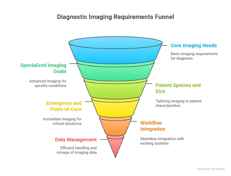
Creating a Diagnostic Requirements Map
A diagnostic requirements map organizes the practice’s needs into categories that correspond to daily clinical use. This provides a clear reference point for comparing equipment options objectively.
A comprehensive map includes:
- Core imaging needs
- Specialized imaging goals
- Patient species and size range
- Emergency and point-of-care requirements
- Workflow integration demands
- Data management expectations
- Long-term expansion considerations
This tool prevents decision fatigue by providing a structured method for comparing machines based on clinical priorities rather than subjective impressions.
Scoring Veterinary Ultrasound Systems Against Clinical Demands
A scoring framework allows for consistent evaluation of multiple systems. Each machine can be rated on resolution, depth penetration, Doppler performance, transducer compatibility, ergonomics, portability, and long-term value.
The scoring system should reflect the diagnostic requirements map, ensuring that essential criteria carry the most weight. This prevents feature overload from skewing the decision toward systems with impressive but unnecessary capabilities.
A structured scoring approach helps the practice align the final choice with real-world clinical needs, supporting a more objective purchase decision.
Why a Clinical-First Framework Leads to Better Outcomes
A clinical-first framework anchors the purchase decision in the realities of daily veterinary practice. It ensures that the machine selected performs reliably across the full range of patient cases, integrates cleanly into workflow, and supports long-term clinical growth.
By organizing the decision process around diagnostic demands, ergonomics, workflow efficiency, and durability, practices avoid common pitfalls such as overemphasizing specifications or focusing solely on price. The result is an ultrasound system that enhances diagnostic capability, supports practice development, and delivers sustained operational value.
FAQ’s
What features matter most in veterinarian ultrasound machines?
The most important features depend on the practice’s diagnostic profile. Resolution, depth penetration, transducer selection, Doppler modes, and ergonomics typically drive clinical performance. Evaluating these elements within real case patterns creates a more accurate assessment than relying on generic specifications.
How should a veterinary practice decide which transducers to purchase?
Clinicians should base transducer selection on the species, case volume, and types of exams performed daily. A combination of microconvex, phased-array, linear, and curvilinear probes often supports a full range of abdominal, cardiac, soft-tissue, and reproductive imaging needs.
Is portability important in vet ultrasound equipment?
Portability matters when the machine is used in multiple rooms, field settings, or emergency contexts. Practices with stable imaging environments may prioritize workstation ergonomics over mobility. The decision depends on how the machine fits into daily workflow.
How long should a veterinarian ultrasound machine last?
High-quality systems typically provide long-term value when maintained correctly. Durability factors such as transducer construction, cable strength, and chassis design influence lifespan more than any specific timeline. Selecting a robust system aligned with clinical needs supports longevity.
Do all veterinary clinics need full Doppler capability?
Not all clinics require advanced Doppler modes, but practices performing cardiac, vascular, or renal assessments benefit strongly from them. Doppler capability should match the complexity of expected diagnostic work.
How does ultrasound equipment affect practice revenue?
Ultrasound increases in-house diagnostic capability, reduces the need for external referrals, and supports expanded clinical services. A machine that delivers reliable image quality and integrates easily into workflow typically supports higher usage and stronger revenue generation.
Can a general practice use the same ultrasound machine for exotic animals?
Some machines provide the resolution required for exotic species, while others do not. Practices treating small mammals, birds, or reptiles often require high-frequency transducers capable of capturing small anatomical structures clearly.
How does workflow influence ultrasound equipment selection?
Workflow determines how frequently the machine is used, how quickly exams must be performed, and how many clinicians share the system. Aligning equipment choices with workflow patterns enhances efficiency and diagnostic consistency.