Point-of-care ultrasound (POCUS) machines have transformed medical diagnostics, enabling real-time imaging at the bedside. These portable devices offer healthcare professionals rapid insights into patient conditions, reducing reliance on traditional imaging methods that often involve delays.
As we move through 2025, several key advancements in POCUS technology are shaping the future of medical imaging. From AI integration to enhanced portability and broader clinical applications, the evolution of these devices is set to further improve diagnostic accuracy and patient care.
Artificial Intelligence and Machine Learning in POCUS
One of the most groundbreaking developments in ultrasound technology is the integration of artificial intelligence (AI) and machine learning. These innovations allow for:
- Automated Image Analysis – AI-powered algorithms can now detect and highlight abnormalities, reducing the margin of error.
- Real-Time Guidance – Intelligent software can assist healthcare providers by offering real-time feedback on probe positioning and image interpretation.
- Workflow Optimization – AI integration streamlines the imaging process, reducing the time needed for manual interpretation and documentation.
These enhancements improve efficiency while enabling even less-experienced users to obtain high-quality ultrasound scans.
The Rise of Handheld and Wearable Ultrasound Devices
Recent advancements in ultrasound technology have led to the development of smaller, more portable devices. Modern POCUS machines are now:
- More Compact and Lightweight – Many models are handheld and can be connected to smartphones or tablets.
- Battery-Powered and Wireless – This eliminates the need for complex setups, making them highly accessible in remote locations.
- More Affordable – Cost reductions have increased accessibility in smaller clinics and underdeveloped regions.
Wearable ultrasound devices are also emerging, providing continuous patient monitoring for conditions such as heart failure and pulmonary edema.
Cloud-Based Connectivity and Telemedicine Integration
Wireless connectivity and cloud-based storage have become essential features in modern POCUS devices. These advancements provide:
- Instant Data Sharing – Clinicians can send ultrasound images to specialists in real time for second opinions.
- Remote Consultations – POCUS is now a valuable tool for telemedicine, allowing providers to assess patients from a distance.
- Seamless Integration with Electronic Health Records (EHRs) – This ensures patient imaging data is accessible across multiple healthcare platforms.
These features are particularly beneficial in rural and underserved areas, where access to medical specialists is limited.
Expansion of Clinical Applications
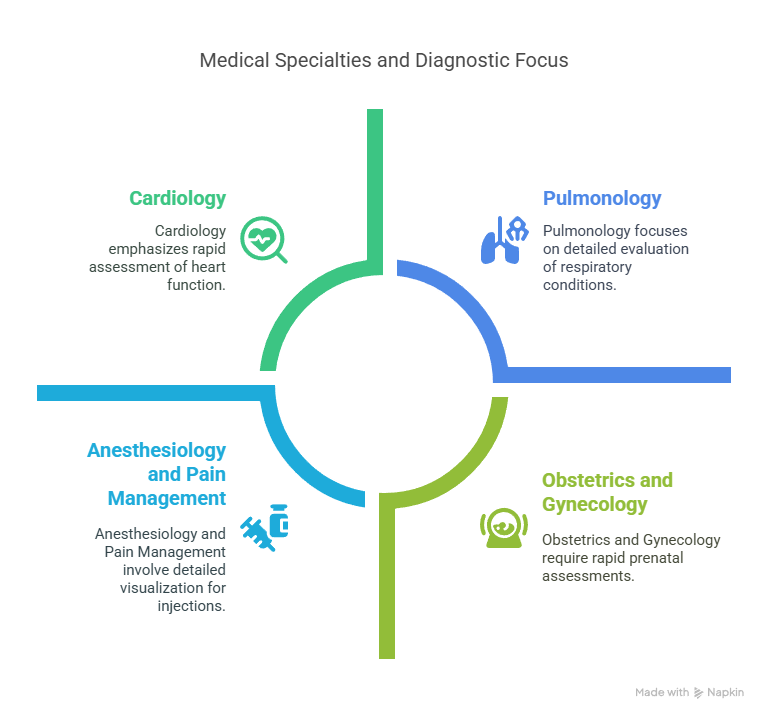
Originally used primarily in emergency and critical care settings, POCUS is now being adopted across various specialties:
- Cardiology – Rapid bedside assessments of heart function, identifying fluid buildup and valve abnormalities.
- Obstetrics and Gynecology – Immediate fetal monitoring, prenatal assessments, and evaluations of reproductive health.
- Pulmonology – Detection of conditions such as pleural effusions, pneumonia, and lung collapse.
- Anesthesiology and Pain Management – Improved visualization for regional anesthesia and pain control injections.
With ongoing advancements, POCUS is expected to play an even greater role in outpatient settings, primary care, and pre-hospital emergency medicine.
High-Frequency and 3D/4D Imaging Enhancements
Modern POCUS machines now offer improved imaging capabilities, including:
- High-Frequency Probes – Providing enhanced resolution for superficial structures such as blood vessels, nerves, and tendons.
- 3D and 4D Ultrasound – Enabling more detailed imaging of organs and fetal development.
- Contrast-Enhanced Ultrasound (CEUS) – Improving the detection of tumors and vascular abnormalities.
These advancements make ultrasound a more powerful diagnostic tool, offering higher clarity and greater diagnostic confidence.
Robotic and Automated Ultrasound Systems
Automation is becoming a major focus in ultrasound technology. Some of the latest developments include:
- Robotic-Assisted Scanning – Reducing user dependency and ensuring consistency in probe positioning.
- Automated Measurements – Eliminating manual calculations for cardiac and fetal assessments.
- Self-Guided Systems – AI-powered ultrasound machines that can autonomously perform scans and provide preliminary findings.
These innovations address the shortage of trained sonographers and improve the overall efficiency of ultrasound examinations.
Focused Ultrasound-Mediated Diagnostics: Enhancing Non-Invasive Disease Detection
Focused ultrasound (FUS) has emerged as a transformative modality in medical diagnostics, offering non-invasive techniques to detect and monitor various diseases. By concentrating ultrasound energy on specific tissues, FUS facilitates precise imaging and therapeutic interventions without the need for surgical procedures.
In Vivo Applications
One notable application of FUS is in the realm of in vivo diagnostics, particularly for blood-related diseases. Techniques such as “sonocytometry” utilize high-intensity focused ultrasound (HIFU) to detect particle sizes within blood flow, enabling the identification of conditions like anemia and leukemia. This method leverages the variability between acoustic impedance to differentiate particles from the medium, providing a non-invasive alternative to traditional blood sampling.
Additionally, photoacoustic (PA) flow cytography has been developed to detect circulating tumor cells (CTCs) in the bloodstream. By targeting the unique absorption spectra of melanoma cells, this technique allows for the identification and immediate eradication of malignant cells through thermal ablation. Such advancements hold promise for early cancer detection and treatment monitoring.
Acoustofluidics for Vasculature Imaging
The integration of acoustofluidics with FUS has opened new avenues for vasculature imaging. This approach employs super-harmonic ultrasound signals to achieve high-resolution images of microvascular structures. When combined with targeted microbubbles, acoustofluidics facilitates detailed three-dimensional imaging of tumor angiogenesis, aiding in the diagnosis and treatment planning for cancers.
Targeted microbubbles, functionalized with specific ligands, have been utilized to detect pathological conditions such as tumor-induced angiogenesis and inflammation. These microbubbles bind to molecular markers like VEGFR2 and αvβ3 integrin, prevalent in tumor vasculature, enabling precise imaging of cancerous tissues. Beyond oncology, molecular ultrasound imaging has been applied to assess inflammation in atherosclerosis, providing insights into plaque buildup and cardiovascular risk.
Challenges and Limitations
Despite the promising advancements, focused ultrasound-mediated diagnostics face several challenges. Manufacturing complexities, including the need for cleanroom environments and specialized materials, hinder large-scale production and portability. Health concerns also arise, as prolonged exposure to FUS at certain wavelengths can induce tissue damage, necessitating the establishment of safety parameters. Furthermore, while in vitro applications of FUS can detect cell size and count, they often lack detailed information about cellular structures or organelles, limiting their diagnostic utility.
Ultrasound-Modulated Optical Tomography: Advancements in Imaging
Ultrasound-modulated optical tomography (UOT) represents a significant leap in imaging technology, combining the high contrast of optical imaging with the spatial resolution of ultrasound. Initially proposed for virus detection, UOT has evolved to enhance imaging depth and resolution through various innovations.
Development and Enhancements
Recent advancements in UOT include the use of coded ultrasound transmissions to improve signal-to-noise ratios, homodyne time-of-flight techniques for better detection sensitivity, and super-resolution methods to surpass traditional imaging limitations. These developments have collectively enhanced the applicability of UOT in clinical diagnostics, offering detailed insights into tissue structures and functions.
Multispectral Optoacoustic Tomography: A Multifaceted Imaging Approach
Multispectral optoacoustic tomography (MSOT) integrates optical and ultrasound imaging to provide real-time, high-resolution images of biological tissues. By detecting ultrasonic waves generated by tissue absorption of pulsed laser light, MSOT offers functional and molecular information beyond conventional imaging modalities.
Emerging Clinical Applications
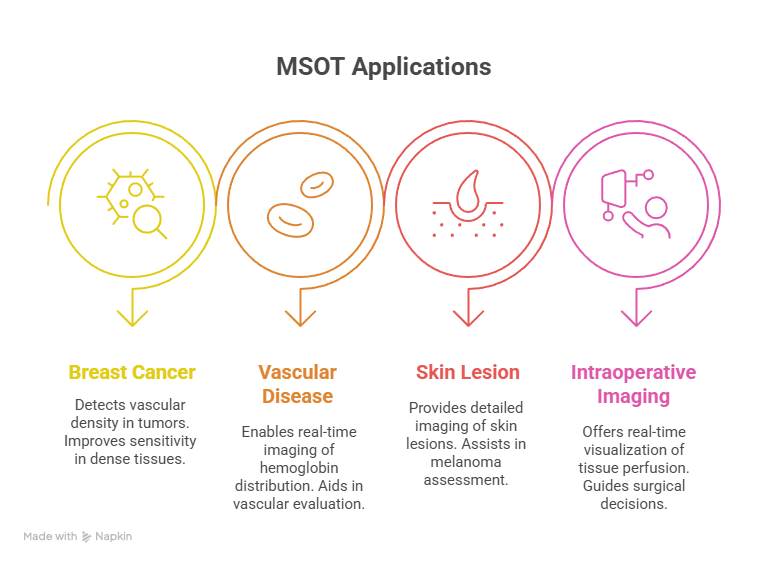
MSOT has shown potential in various clinical applications:
- Breast Cancer Imaging: MSOT can detect increased vascular density associated with tumors, offering improved sensitivity over traditional mammography, especially in dense breast tissues.
- Vascular Disease Assessment: The technology enables real-time imaging of hemoglobin distribution in carotid arteries, aiding in the evaluation of vascular diseases.
- Skin Lesion Evaluation: MSOT provides detailed imaging of skin lesions and their vascular networks, assisting in the assessment of conditions like melanoma.
- Intraoperative Imaging: During surgeries, MSOT offers real-time visualization of tissue perfusion, helping to identify areas at risk of ischemia and guiding surgical decisions.
Integration with Point-of-Care Ultrasound Systems
The integration of advanced imaging techniques like MSOT with point-of-care ultrasound systems enhances diagnostic capabilities at the bedside. This fusion allows clinicians to obtain comprehensive information about tissue morphology and function without the need for invasive procedures, thereby improving patient outcomes and expanding the utility of POCUS devices.
Focused Ultrasound in Neurological Applications
Focused ultrasound is making significant strides in neurology, particularly in the modulation of brain activity. Innovative brain-computer interfaces employing ultrasound are under investigation to enhance mood and treat conditions such as depression, addiction, obsessive-compulsive disorder, and epilepsy. By precisely targeting neuron clusters, these non-invasive techniques aim to correct imbalanced brain activity patterns, offering new therapeutic avenues for neurological disorders.
Artificial Intelligence in Ultrasound Diagnostics
The integration of artificial intelligence (AI) into ultrasound diagnostics is transforming disease detection and management. For instance, AI models have been developed to diagnose pneumonia, COVID-19, and other lung diseases from ultrasound videos with remarkable accuracy. These models analyze video frames for specific lung features and patterns, classifying the findings into diagnostic categories. The use of explainable AI techniques ensures that radiologists can comprehend and trust the model’s decisions, thereby enhancing diagnostic speed, accuracy, and serving as valuable training tools for clinicians.
Networked Medical Devices and the Medical Internet of Things (MIoT)
The integration of networked medical devices, including ultrasound machines, ventilators, and data-sharing implants, is transforming patient care and medical research. The Medical Internet of Things (MIoT) market is experiencing significant growth, driven by innovations that enable early detection of medical conditions and improved post-operative monitoring. These technologies leverage data analytics, cloud computing, and AI to provide more objective and efficient medical assessments. However, increased connectivity also raises cybersecurity concerns
Training and Education in POCUS
With the increasing use of ultrasound, healthcare professionals require proper training to maximize the benefits of POCUS. Some key advancements in training include:
- Virtual Reality (VR) and Augmented Reality (AR) Simulations – Offering immersive learning experiences for trainees.
- Online Certification Programs – Allowing more medical professionals to become proficient in ultrasound use.
- AI-Guided Training Modules – Providing real-time feedback during practice sessions.
These educational tools ensure that POCUS remains an accessible and effective diagnostic modality across all healthcare levels.
Market Growth and Global Accessibility
The demand for POCUS is rapidly increasing due to its versatility, affordability, and ease of use. Key factors driving this growth include:
- Affordability and Cost-Effectiveness – POCUS devices are more affordable than traditional imaging systems like MRI and CT scans.
- Increased Adoption in Low-Resource Settings – Portable ultrasound machines are being used in rural clinics and developing countries.
- Government and Institutional Support – Many healthcare organizations are investing in POCUS to improve patient outcomes.
As technology continues to evolve, the widespread adoption of POCUS is expected to bridge gaps in medical imaging accessibility worldwide.
Looking Ahead: The Future of POCUS
The next decade will see even more innovations in ultrasound technology. Trends expected to shape the future include:
- Wearable and AI-Powered Devices – Continuous real-time monitoring of at-risk patients.
- Improved Battery Life and Energy Efficiency – Allowing longer use in field and emergency settings.
- More Advanced Imaging Modalities – Including holographic ultrasound for enhanced 3D visualization.
With ongoing research and technological advancements, POCUS will continue to be a vital tool in modern medicine, further transforming how healthcare professionals diagnose and treat patients.
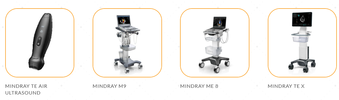
Highlighted Mindray point-of-care ultrasound machine models available through BOMImed include Mindray TE Air Ultrasound, Mindray M9, Mindray ME 8, Mindray TE X, Mindray TE5 SP, Mindray TE7 Max, Mindray Z.One Pro. Discover the difference with BOMImed’s comprehensive range of portable ultrasound machines, designed to enhance diagnostic efficiency and patient care.
Shop by category: Point of Care Ultrasound – Portable Ultrasound – Radiology Ultrasound