In medical imaging, ultrasound transducers are integral to obtaining high-quality diagnostic images. An ultrasound transducer is a critical component in ultrasound systems, converting electrical energy into sound waves that penetrate the body and reflect back to create images of tissues and organs. Understanding the basics of how ultrasound transducers work and their various types can significantly enhance one’s grasp of their application in modern medical diagnostics.
How Does an Ultrasound Transducer Work?
Ultrasound transducers operate on the principle of piezoelectricity, where specific materials generate sound waves when subjected to an electric field. This process begins with the transducer’s crystal elements, which vibrate upon receiving electrical pulses. These vibrations produce sound waves that travel through the body and reflect off tissues. The transducer then captures the returning echoes, converting them back into electrical signals that are processed to form an image.
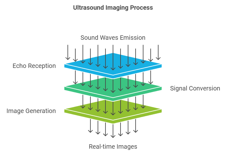
Steps in the Ultrasound Imaging Process
- Transmission of Sound Waves: The transducer emits sound waves that travel through the body until they hit boundaries between different tissues.
- Reception of Echoes: When the sound waves hit an organ or tissue boundary, they bounce back toward the transducer.
- Signal Conversion: The transducer detects these returning echoes and converts them into electrical signals.
- Image Generation: The ultrasound system processes these signals to produce real-time images, allowing practitioners to examine internal structures non-invasively.
For a more in-depth understanding, consider learning about how ultrasound machines work.
Types of Ultrasound Transducers
Various types of ultrasound transducers are tailored to specific applications, from imaging deep organs to visualizing superficial structures. Here are some common types:
- Linear Transducers: Ideal for imaging superficial structures such as muscles, tendons, and thyroids.
- Curved Array Transducers: Useful for abdominal scans, as their design allows for deeper penetration.
- Phased Array Transducers: Often employed in cardiac imaging because of their narrow field and ability to fit between the ribs. This phased array ultrasound transducer is also used for scanning areas where space is limited.
Each transducer type differs in design and frequency range, allowing specialists to select the most suitable option for each diagnostic need.
RELATED: Choosing the Right Portable Ultrasound Machine
Frequency and Resolution in Ultrasound Transducers
The frequency of the ultrasound transducer plays a significant role in determining image clarity and depth. Higher frequencies provide greater resolution but have limited penetration, making them ideal for shallow examinations. Lower frequencies, in contrast, offer deeper penetration at the cost of resolution. This frequency selection process is crucial in diagnostic ultrasound to ensure images are both clear and accurate.
Care and Maintenance of Ultrasound Transducers
Maintaining transducers is vital for both performance and patient safety. Proper handling and cleaning are essential to extend the transducer’s lifespan and ensure hygienic conditions.
Cleaning Protocols
- Use of Approved Disinfectants: After each use, transducers should be cleaned with disinfectants approved by the manufacturer to prevent cross-contamination and maintain functionality.
- Avoiding Abrasive Materials: Harsh chemicals and abrasive materials can damage the transducer’s surface.
- Routine Checks: Regular inspection of the transducer’s surface and cable can help identify potential issues early on.
For guidance on proper maintenance, review technical support for ultrasound equipment.
How to Hold an Ultrasound Transducer
The way a transducer is held affects image quality and the accuracy of results. Training in how to hold an ultrasound transducer helps technicians and healthcare professionals gain proficiency, ensuring they capture the best possible images. Proper hand positioning can reduce strain and improve the accuracy of the scan, particularly during extended procedures.
Costs Associated with Ultrasound Transducers
The cost of an ultrasound transducer varies widely depending on the type, application, and manufacturer. High-frequency transducers or those designed for specific imaging purposes tend to be more expensive due to their specialized construction. Costs can range from several hundred to thousands of dollars. Decision-makers investing in ultrasound equipment should assess the balance between performance needs and budget constraints.
Key Components and Design of an Ultrasound Transducer
Understanding the core design elements of an ultrasound transducer can provide insights into its functionality and importance. The primary parts of a transducer include:
- Piezoelectric Crystals: The most essential component, these crystals are responsible for generating sound waves when an electric current is applied and for receiving echoes from tissues.
- Backing Material: Located behind the piezoelectric element, this material dampens the vibrations to ensure precise sound wave generation and reduce noise.
- Matching Layer: Positioned between the piezoelectric element and the patient’s skin, this layer minimizes impedance mismatch, enhancing the transmission of sound waves.
- Acoustic Lens: This component focuses the sound waves, improving image quality and resolution.
Together, these elements allow the transducer to generate and capture the echoes needed for high-quality imaging. The careful engineering of each part determines how well an ultrasound transducer can perform across various applications.
Different Types of Ultrasound Transducers and Their Applications
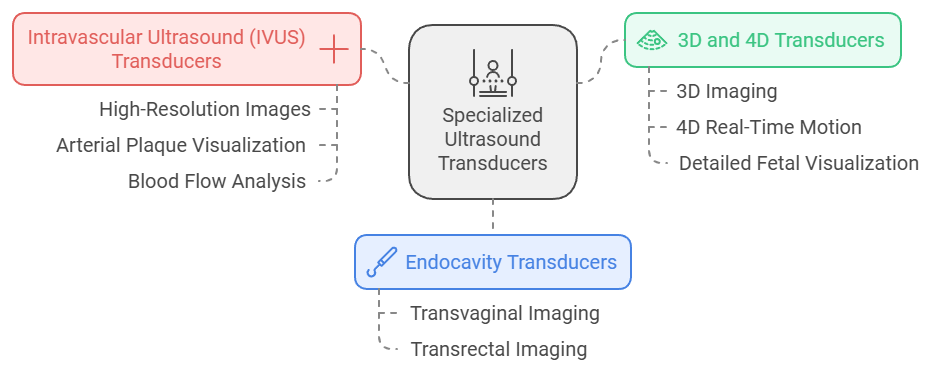
Each type of ultrasound transducer is designed with specific imaging needs in mind. Here are some specialized transducers and their applications:
- Endocavity Transducers: Used for internal examinations such as transvaginal and transrectal imaging, these transducers are designed to fit within body cavities, allowing for close-range imaging.
- Intravascular Ultrasound (IVUS) Transducers: IVUS transducers are miniature devices inserted into blood vessels to produce high-resolution images from inside the artery, essential in cardiology for visualizing arterial plaque and blood flow.
- 3D and 4D Transducers: Used in obstetric imaging, these transducers capture three-dimensional images, while 4D imaging adds real-time motion, enabling detailed visualization of a fetus.
For a comprehensive look at ultrasound technology applications, refer to understanding ultrasound imaging technology.
How Transducer Frequency Affects Imaging
The frequency of an ultrasound transducer affects not only image quality but also the types of examinations it can perform. Here’s a breakdown of how frequency influences imaging:
- High-Frequency Transducers (10-15 MHz): These transducers offer excellent resolution for imaging shallow structures, like the skin and muscles, but they cannot penetrate deep into the body.
- Mid-Frequency Transducers (5-10 MHz): Commonly used for abdominal imaging, this range balances penetration depth with image clarity.
- Low-Frequency Transducers (2-5 MHz): Suitable for deeper examinations, such as cardiac or abdominal imaging, though lower frequencies typically provide less detail.
Medical professionals must select the transducer frequency based on the specific diagnostic requirements. For example, a cardiologist might use a low-frequency phased array transducer to examine heart chambers, while a dermatologist would choose a high-frequency linear transducer to assess skin structures.
RELATED: Introduction to Point-of-Care Ultrasound Machines
Advanced Imaging Techniques with Phased Array Transducers
The phased array ultrasound transducer is widely used in cardiology, emergency medicine, and other fields requiring compact, precise imaging. This transducer type uses multiple piezoelectric elements activated in rapid sequences, allowing sound waves to steer and focus electronically. Phased array transducers can:
- Capture Real-Time Heart Function: By producing high-quality images through small acoustic windows, these transducers allow for detailed visualization of heart chambers and valves.
- Examine Small, Hard-to-Reach Areas: The narrow footprint of phased array transducers makes them ideal for imaging areas between the ribs or small body parts.
- Facilitate Point-of-Care Ultrasound (POCUS): Compact and versatile, phased array transducers enable on-the-spot diagnostics, which is essential in emergency and critical care settings.
Explore the benefits of point-of-care ultrasound in medical practice to understand how such innovations streamline clinical workflows.
Proper Maintenance to Extend the Life of Ultrasound Transducers
Investing in high-quality transducers can yield exceptional imaging results, but protecting that investment requires careful maintenance and handling. Here’s a guide to maintaining transducers:
- Routine Cleaning and Disinfection: After each use, clean the transducer with an approved disinfectant to prevent infection and maintain clear imaging. Proper disinfection protocols are particularly critical for endocavity and invasive transducers.
- Inspection for Damage: Regularly inspect the transducer for any cracks, fraying cables, or other signs of wear. Even minor defects can impact image quality and increase patient risk.
- Storage in Protective Cases: Always store transducers in protective cases when not in use to prevent physical damage.
- Avoiding Excessive Heat: Exposure to high temperatures can damage the transducer crystals and internal components, so avoid heat sources during cleaning or storage.
For information on safe handling and equipment care, consult clinical support resources for ultrasound professionals.
Practical Tips on Holding and Using an Ultrasound Transducer
Correct handling of the transducer ensures optimal image quality and can reduce user fatigue. Key tips include:
- Positioning for Image Clarity: Align the transducer carefully and use steady pressure to maintain contact with the skin or surface being imaged.
- Rotating for Different Views: Slightly rotate or tilt the transducer to achieve multiple views without repositioning the patient, which is especially helpful in cardiac and abdominal imaging.
- Reducing Strain: Ergonomic grips and gentle wrist movement can reduce strain on the operator’s hand, which is essential for extended procedures.
Practitioners who become proficient in how to hold an ultrasound transducer can conduct more efficient and accurate scans, improving patient comfort and reducing exam times.
Innovations in Portable and Handheld Ultrasound Transducers
With advancements in portable ultrasound machines, ultrasound imaging is now accessible beyond traditional healthcare facilities. Portable and handheld transducers offer several advantages:
- Greater Accessibility: Medical professionals can perform diagnostics directly at the patient’s bedside, in ambulances, or even in remote locations.
- Reduced Costs: Portable devices can be more cost-effective than full-size ultrasound systems, making ultrasound technology more accessible to clinics and smaller practices.
- Enhanced Flexibility: Handheld ultrasound systems provide on-the-go imaging, ideal for emergency responders, sports medicine, and rural healthcare settings.
Learn more about portable ultrasound machines and how they are transforming healthcare delivery by offering flexibility and faster diagnosis.
Assessing Ultrasound Transducer Costs and Investment Decisions
Purchasing ultrasound equipment, particularly ultrasound transducers, requires a well-informed investment strategy. Costs vary depending on transducer type, imaging quality, and additional features. Here’s what impacts transducer pricing:
- Type and Specialization: High-frequency and specialized transducers, such as 3D/4D or phased array, are typically more expensive due to their complex design.
- Brand and Quality: Trusted brands known for high performance may charge a premium, but they often deliver longer-lasting, reliable products.
- Additional Capabilities: Transducers with advanced features, like Doppler imaging for blood flow analysis, generally cost more due to enhanced technology.
Decision-makers must balance their budget with clinical requirements to make an informed investment. For further guidance on procurement and financing, access service and training resources for ultrasound equipment.
Why Ultrasound Transducers are Vital in Modern Healthcare
Ultrasound transducers play an essential role in diagnostics by enabling non-invasive, real-time imaging. From their intricate piezoelectric technology to advanced models like the phased array transducer, these devices support diverse clinical needs across cardiology, obstetrics, and emergency care. Proper care and routine maintenance ensure their longevity and performance, while innovations in portable ultrasound systems are expanding the reach and effectiveness of ultrasound diagnostics.
Whether it’s understanding how ultrasound transducer frequency impacts imaging, learning to handle these devices proficiently, or staying updated on emerging technologies, having a foundational knowledge of ultrasound transducers empowers medical professionals and stakeholders to make informed, impactful decisions in patient care.


