Point of Care Ultrasound (POCUS) refers to the use of portable ultrasound at a patient’s bedside for diagnostic and therapeutic purposes. This technology allows for immediate visualization of organ systems and is used across various medical fields, revolutionizing the way care is delivered, particularly in emergency and critical care settings.
The Evolution of POCUS
History of Ultrasound in Medicine
The history of ultrasound in medicine is a fascinating journey of innovation and practical application. It began in the early 20th century, but significant development occurred post-World War II, thanks to advancements in sonar technology used during the war. The first practical application of ultrasound in medicine was in 1942 by neurologist Karl Dussik of Austria, who attempted to locate brain tumors and cerebral ventricles by transmitting ultrasound beams through the human skull.
In the 1950s, George Ludwig at the Naval Medical Research Institute used ultrasound to detect gallstones, and in 1956, Ian Donald and his colleagues at the University of Glasgow developed the prototype of an ultrasound machine that could be used for obstetric purposes. This period marked the start of detailed ultrasound research and its application in different fields of medicine.
Throughout the 1960s and 1970s, technological advancements in ultrasound improved image quality, which allowed for its broader application in medical diagnostics. Real-time scanners in the late 1970s and the introduction of Doppler technology further expanded its capabilities, enabling not just structural imaging but also the assessment of blood flow and cardiac activity. By the end of the 20th century, ultrasound had become a crucial tool in cardiology, obstetrics, and radiology, thanks to continuous improvements in transducer technology and image processing.
Emergence of POCUS
The concept of Point of Care Ultrasound (POCUS) emerged from the need to make ultrasound technology more accessible and to provide immediate diagnostic insights that could be critical in emergency situations. The traditional use of ultrasound involved large, immobile machines that were primarily located in radiology departments. This setup was not conducive to situations where quick decision-making based on immediate diagnostic data was crucial, such as in emergency rooms or in intensive care units.
The shift towards POCUS began in the 1990s, as smaller, portable ultrasound devices became available. These devices made it feasible to bring ultrasound directly to the bedside, thereby significantly cutting down the time needed for diagnosis and treatment. Dr. Harvey L. Nisenbaum, a key advocate for the early adoption of POCUS, played a significant role in promoting its use in emergency settings, highlighting its ability to rapidly differentiate between life-threatening conditions and more benign cases.
Another pivotal development was the endorsement of POCUS by various medical bodies. For instance, the American College of Emergency Physicians (ACEP) established guidelines in 2001 for the use of POCUS in emergency medicine, which bolstered its acceptance across various medical specialties. This endorsement was supported by a growing body of research that underscored the benefits of POCUS, including faster diagnostic times and improved patient outcomes.
Technological Advances
The advancement of POCUS is tightly linked to technological progress in ultrasound imaging systems. Early portable ultrasound machines, although less cumbersome than their predecessors, still had limitations in terms of image quality and battery life. However, the 21st century ushered in a new era of innovation, characterized by significant improvements in these areas.
 The introduction of hand-carried ultrasound (HCU) devices in the early 2000s was a game-changer. These devices were not only portable but also provided a level of image quality that was close to that of the larger, more traditional systems. Companies like Sonosite and GE Healthcare were pioneers in this space, developing machines that were robust yet compact enough to be easily transported and used in tight spaces typical of emergency settings.
Further enhancements included the development of wireless transducers and the integration of ultrasound software with smartphones and tablets, expanding the usability and functionality of POCUS systems. The most recent innovations involve the use of artificial intelligence to assist in image interpretation, which helps to reduce the variability in diagnosis associated with operator skill level.
Technical Aspects of POCUS
Equipment and Tools
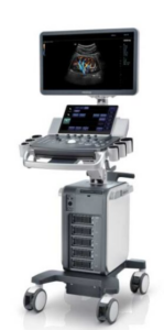 Point of Care Ultrasound (POCUS) utilizes specialized equipment that is designed for portability, ease of use, and rapid deployment in a variety of medical settings. The core components of a POCUS system include the ultrasound machine itself, which is typically compact and battery-operated, and a range of transducers (also known as probes), each suited to different diagnostic tasks.
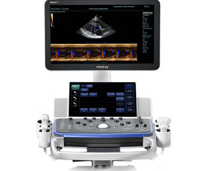
Ultrasound Machines: Modern POCUS machines are remarkably smaller and lighter than their traditional counterparts, which allows them to be easily transported to patient bedsides, emergency scenes, or in field settings. These machines are equipped with high-resolution screens and user-friendly interfaces that simplify the process of acquiring and interpreting images. Some models are even integrated into handheld devices or connect to tablets, further enhancing their portability and convenience.
Transducers: POCUS systems use various types of transducers to capture images of different parts of the body. The choice of transducer depends on the depth and nature of the tissue being examined:
- Linear probes are used for superficial structures such as blood vessels, muscles, and nerves.
- Curved probes provide broader and deeper penetration, suitable for abdominal and obstetric imaging.
- Phased array probes are designed for cardiac imaging, capable of capturing deeper structures through a smaller acoustic window.
- Software and Image Processing: Advanced software algorithms play a crucial role in optimizing image quality and facilitating interpretation. Features like color Doppler can be used to assess blood flow, while newer technologies incorporate machine learning algorithms to aid in identifying structures and pathologies, making it easier for clinicians with varying levels of ultrasound training to use POCUS effectively.
Performing a POCUS Examination
Conducting a POCUS examination involves several steps that ensure accurate and effective imaging. The procedure typically follows this sequence:
Patient Preparation: Ensure the patient is positioned comfortably and appropriately based on the area to be examined. For example, for cardiac imaging, the patient might be positioned in the left lateral decubitus position.
Machine Setup: Power on the ultrasound machine and select the appropriate settings and probe based on the examination objectives. Adjust the depth, gain, and focus to optimize image quality.
Probe Placement: Apply a generous amount of ultrasound gel to the probe to facilitate smooth contact with the skin and eliminate air pockets, which can interfere with sound wave transmission. Position the probe correctly, starting with a standard orientation and adjusting based on the anatomical structures and patient specifics.
Image Acquisition: Move the probe along the area of interest to explore different angles and depths. Systematically cover the area to ensure no pathologies are missed. Utilize different modes such as B-mode for structural assessment and Doppler modes for evaluating blood flow.
Interpretation: Analyze the images in real time to make clinical decisions. Look for normal anatomical structures and signs of any abnormalities.
Documentation: Save important images and clips within the patient’s medical record for future reference and for discussion with other healthcare professionals.
Training and Competency
Proper training is crucial to the effective use of POCUS, as the interpretation of ultrasound images requires both technical skill and clinical knowledge. Training typically includes:
 Basic Training: Introductory courses that cover the fundamentals of ultrasound physics, instrument handling, and basic scanning techniques. These are often provided by hospitals, academic institutions, or through specialty organizations.
Specialized Training: Advanced courses focusing on specific applications of POCUS, such as emergency medicine, critical care, or cardiology. These courses may include hands-on sessions, simulations, and supervised clinical practice.
Certification and Continuing Education: Various professional bodies offer certification programs that validate a practitioner’s skills in POCUS. Maintaining competency involves continuous education and practice, staying updated with the latest advancements and best practices in ultrasound technology.
The technical aspects of POCUS underscore its adaptability and utility in modern medicine, where rapid, non-invasive, and accurate diagnostic capabilities are paramount. As technology evolves, so too will the training and tools associated with POCUS, making it an increasingly indispensable part of medical care across specialties.
Applications of POCUS
Point of Care Ultrasound (POCUS) has revolutionized the speed and manner in which medical care is delivered, particularly in acute and critical care settings. Its versatility allows for immediate diagnostic insights across a variety of specialties, enhancing patient management and outcomes.
Emergency Medicine
In emergency medicine, POCUS is a critical tool that provides rapid diagnostics crucial in life-saving situations. Its ability to be used at the bedside or in the field makes it especially valuable in trauma care, where time-sensitive decisions can dramatically affect outcomes.
FAST Exam: The Focused Assessment with Sonography for Trauma (FAST) exam is one of the most common applications of POCUS in emergency settings. It is designed to quickly identify free fluid (blood) in the abdominal cavity, which can indicate internal bleeding. The FAST exam has significantly improved the speed at which life-threatening injuries are identified in trauma patients.
Cardiac Evaluation: POCUS is used to assess cardiac function in emergency settings, providing immediate information about cardiac activity, which is crucial for patients presenting with chest pain or symptoms of heart failure. It allows for the rapid assessment of ejection fraction, cardiac tamponade, and other conditions that may require urgent intervention.
Lung and Pleural Assessment: POCUS helps in the evaluation of respiratory distress by identifying pneumothorax, pleural effusions, and pulmonary edema. This is particularly useful in settings where X-ray or CT imaging is not immediately available.
Vascular Access: POCUS enhances the success rate and safety of procedures like central venous catheterization by providing real-time imaging that guides needle placement, reducing the risk of complications such as arterial puncture or hematoma.
Critical Care
 In critical care, the role of POCUS extends to almost continuous monitoring and diagnostic evaluations due to the dynamic nature of patient status in intensive care units (ICUs).
Hemodynamic Monitoring: POCUS is invaluable for non-invasive hemodynamic monitoring, assessing parameters such as preload, cardiac output, and fluid responsiveness. This is crucial in the management of shock and sepsis, where fluid status and cardiac function are continuously in flux.
Renal and Bladder Function: It assists in assessing bladder volume for urinary retention and can evaluate kidney size and structure to aid in diagnosing acute kidney injury or chronic kidney disease.
Neurological Applications: While still in developmental stages, POCUS is being used to assess optic nerve diameter, which can correlate with intracranial pressure, an important consideration in patients with head injuries or neurological disorders.
Other Specialties
The flexibility of POCUS allows it to be adapted across various other medical fields, each benefiting from the specific capabilities of ultrasound technology.
Obstetrics and Gynecology: POCUS facilitates immediate visualization of the fetus and uterus, aiding in the assessment of fetal health, placental location, and amniotic fluid volume. It is also used in gynecological emergencies to assess for conditions like ectopic pregnancy and ovarian torsion.
Pediatrics: Safe and non-invasive, POCUS is ideal for pediatric care, used for assessing hip dysplasia in infants, diagnosing appendicitis in children, and evaluating for pyloric stenosis.
Rheumatology: POCUS can assess joint spaces and musculoskeletal structures, aiding in the diagnosis and management of inflammatory and non-inflammatory conditions, such as rheumatoid arthritis and gout.
Anesthesiology: Increasingly used in regional anesthesia, POCUS improves the accuracy of nerve blocks and spinal anesthesia applications, enhancing patient safety and the effectiveness of pain management strategies.
Clinical Benefits of POCUS
The clinical benefits of POCUS are extensive, given its non-invasive nature, cost-effectiveness, and ability to provide rapid diagnostic information. Some of the key advantages include:
Improved Diagnostic Accuracy: POCUS offers real-time imaging, improving the accuracy and speed of diagnoses, particularly in emergency and critical care settings where traditional imaging modalities may be too slow or unavailable.
Enhanced Patient Safety: By reducing the need for more invasive diagnostic procedures, POCUS decreases the risk of complications, such as infections or bleeding associated with traditional methods.
Cost-effectiveness: POCUS can reduce the need for expensive diagnostic tests and shorten hospital stays by accelerating decision-making processes, which lowers overall healthcare costs.
POCUS has become an indispensable tool across various medical specialties, transforming the approach to diagnosis and treatment in modern healthcare. Its continued adoption and integration into standard medical protocols underscore its significance in improving patient outcomes and advancing medical care. As technology and training continue to evolve, the applications and benefits of POCUS are likely to expand even further, making it an even more valuable resource in the medical field.
Clinical Benefits of POCUS
Point of Care Ultrasound (POCUS) has emerged as a transformative tool in medicine, offering a range of benefits that enhance clinical outcomes and streamline patient care. Its capacity for real-time, bedside imaging provides critical insights that can influence treatment decisions across various medical disciplines.
Improving Diagnostic Accuracy
One of the paramount benefits of POCUS is its ability to enhance diagnostic accuracy. Its real-time imaging capability allows clinicians to obtain immediate visual insights into the patient’s condition, which is particularly valuable in acute and emergency care settings.
 Rapid Decision-Making: POCUS enables quick diagnoses, often within minutes. This is crucial in situations like trauma, where the FAST (Focused Assessment with Sonography in Trauma) exam can rapidly detect internal bleeding, or in cardiac emergencies where echocardiography can assess heart function on the spot.
Reduction in Diagnostic Uncertainty: Traditional diagnostic tools such as X-rays or CT scans often require transferring the patient to specialized departments, which can delay diagnosis and treatment. POCUS eliminates many of these delays, reducing the window of diagnostic uncertainty and facilitating quicker interventions.
Enhanced Sensitivity and Specificity: In many cases, POCUS can offer comparable, and sometimes superior, sensitivity and specificity to traditional imaging methods. For example, in detecting pneumothorax, POCUS has been shown to outperform chest X-ray in terms of sensitivity, providing more accurate and timely diagnoses.
Enhancing Patient Safety
The use of POCUS significantly enhances patient safety by reducing the need for more invasive, higher-risk procedures and by offering a non-ionizing alternative to radiographic assessments.
Lower Complication Rates: Procedures such as central line placements, thoracenteses, or paracenteses are safer when performed under ultrasound guidance, which reduces the risk of accidental organ puncture or other complications associated with blind insertion techniques.
Non-Ionizing: Unlike CT scans and other radiographic methods, ultrasound does not expose patients to ionizing radiation, making it a safer choice for repetitive use and for populations particularly sensitive to radiation, such as pregnant women and children.
Immediate Troubleshooting: POCUS allows for immediate reassessment of treatment efficacy. For instance, in the case of cardiac tamponade, POCUS can quickly confirm whether pericardiocentesis has successfully relieved the pressure on the heart, allowing for rapid adjustments in care.
Cost-effectiveness
POCUS not only improves the quality of care but also offers significant cost benefits. By integrating POCUS into routine clinical practice, healthcare facilities can achieve substantial savings.
Reduced Need for Expensive Testing: By providing immediate insights, POCUS can often obviate the need for more expensive diagnostic tests. In many cases, a POCUS examination can prevent an unnecessary CT scan or MRI, significantly reducing healthcare costs.
Decreased Hospital Stays: POCUS can expedite diagnosis and the initiation of treatment, potentially reducing the length of hospital stays. For example, early diagnosis of heart failure or abdominal free fluid can lead to quicker management decisions, which can shorten the time a patient spends in the hospital.
Increased Throughput in Emergency Departments: The rapid diagnostic capabilities of POCUS can increase patient throughput in busy settings like emergency departments, improving the efficiency of care delivery and decreasing waiting times for patients.
Broader Access to Diagnostic Tools
The portability and decreasing cost of ultrasound machines have broadened access to diagnostic tools, especially in resource-limited settings or in rural areas where large, expensive imaging equipment is not feasible.
Rural and Remote Areas: In regions with limited medical infrastructure, POCUS provides essential diagnostic capabilities. Its ease of use and portability make it ideal for rural healthcare providers, who can perform scans and obtain immediate results, improving the standard of care in underserved areas.
Global Health Applications: POCUS is increasingly recognized as a vital tool in global health efforts, providing diagnostic capacity in parts of the world where healthcare resources are scarce. Its role in infectious disease management, maternal health, and emergency care is particularly impactful in improving health outcomes in developing countries.
The integration of POCUS into clinical practice represents a significant advancement in the way care is delivered. By improving diagnostic accuracy, enhancing patient safety, and providing cost-effective care, POCUS has established itself as an essential component of modern medicine. Its continued adoption and the ongoing development of its applications promise to further expand its role, making healthcare more accessible and effective worldwide. As technology continues to advance, the potential for POCUS to impact all areas of medicine only increases, reinforcing its value in improving patient outcomes and the efficiency of medical services.
Challenges and Limitations
While Point of Care Ultrasound (POCUS) offers substantial benefits and has been increasingly integrated into various medical practices, it is not without challenges and limitations. Understanding these issues is crucial for optimizing the use of POCUS, enhancing its effectiveness, and addressing potential drawbacks in clinical settings.
Technical Limitations
POCUS is highly operator-dependent, and its effectiveness can vary significantly based on the skill and experience of the user. This variability introduces several challenges:
Image Quality and Interpretation: Unlike more static imaging modalities such as CT or MRI, the quality of ultrasound images is highly dependent on the operator’s technique. Factors such as probe handling, angle, and pressure can substantially affect image clarity and accuracy. Moreover, interpreting ultrasound images requires a deep understanding of anatomical variations and the nuances of ultrasound physics, which can be a significant hurdle for less experienced practitioners.
Equipment Variability: The range of devices available for POCUS use varies widely in terms of quality, capabilities, and ease of use. Lower-cost or older models may lack advanced imaging features such as high-resolution displays or specialized software that aids in interpretation, potentially compromising diagnostic accuracy in less than ideal circumstances.
Training and Standardization
The effectiveness of POCUS is contingent upon adequate training and the standardization of both practice and education. These aspects present their own set of challenges:
Inconsistent Training: There is no universal standard for POCUS training, resulting in variability in skill levels among practitioners. Some clinicians may receive extensive hands-on training, while others might rely on self-taught methods or brief instructional courses. This inconsistency can lead to disparities in the quality of care provided.
Need for Ongoing Education: Ultrasound technology and its applications are continually evolving, necessitating ongoing education and practice to maintain proficiency. Keeping up with the latest techniques and clinical evidence can be challenging for healthcare providers who must balance this need with their clinical duties.
Credentialing and Privileging Issues: Establishing credentials for POCUS usage and determining privileges within institutions can be complex. Hospitals and medical boards must develop criteria that ensure competence without restricting access to this valuable tool unnecessarily.
Ethical and Legal Considerations
The immediate availability of diagnostic information through POCUS can also raise ethical and legal issues that must be carefully managed:
Diagnostic Overshadowing: The rapid insights provided by POCUS can sometimes lead to premature conclusions, potentially overshadowing other diagnostic possibilities and leading to errors in patient management.
Liability Concerns: As with any diagnostic tool, there is a risk of misdiagnosis with POCUS. Given the operator-dependent nature of ultrasound, this risk may be heightened, potentially increasing liability for clinicians and institutions, especially if practitioners are inadequately trained.
Patient Consent and Information: The ease and speed of using POCUS can sometimes bypass standard processes for patient consent and thorough explanation of procedures. Clinicians must remain diligent about informing patients and ensuring their understanding and consent, even in fast-paced environments.
Future Prospects
Despite these challenges, the future of POCUS looks promising, with potential advancements likely to mitigate many current limitations:
Technological Improvements: Ongoing advances in ultrasound technology, such as enhanced image quality, artificial intelligence integration, and more intuitive user interfaces, are likely to reduce the learning curve and improve diagnostic accuracy.
Standardization and Certification: Efforts are underway to standardize training and certification for POCUS, which will help ensure a high level of proficiency across medical professions. Professional organizations and accrediting bodies are increasingly focusing on developing clear guidelines and competency standards.
Expanding Applications: Research continues to expand the applications of POCUS, exploring new ways it can be used in medical diagnostics and treatment. As evidence grows, so too will the confidence in its capabilities, potentially leading to broader acceptance and utilization.
The challenges and limitations of POCUS are significant, yet they are counterbalanced by the enormous benefits and potential that this technology offers to the field of medicine. By addressing these issues through improved training, technological advances, and thoughtful integration into clinical practice, POCUS can continue to evolve as an essential tool in modern healthcare, enhancing patient care and outcomes across a wide range of medical specialties.


